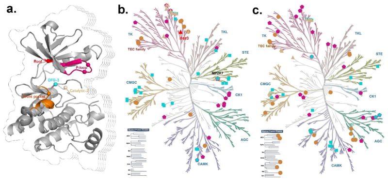
Figure 7.
Reactive cysteines across the human kinome. (a) The reactive cysteines distributed at the five regions of binding sites marked in different colors. (b) The kinases with released 3D kinase structures. (c) The kinases without released kinase structures. (b) and (c) were generated using KinMap (http://kinhub.org/) and the high resolution figures are available in supporting information Figure S2–3.

