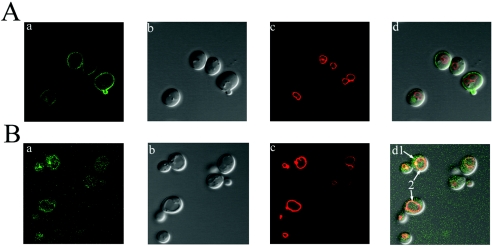
FIG. 2.
Localization of Ist2p to the plasma membrane and alteration of the localization of Ist2p in btn2Δ strains. (A) EGFP-Ist2p in ist2Δ strain B-14915. (B) EGFP-Ist2p in btn2Δ ist2Δ strain B-14983. (a) EGFP fluorescence. (b) Differential interference contrast images. (c) FM4-64 staining showing the vacuolar membrane. (d) Merged images. EGFP-Ist2p in the ist2Δ strain localizes to the plasma membrane. EGFP-Ist2p in the btn2Δ ist2Δ strain appears to be mislocalized to the cytoplasm and the vacuolar membrane (arrows 1 and 2, respectively). Each image is presented at a magnification of ×100 and is typical of that seen for the entire cell population.

