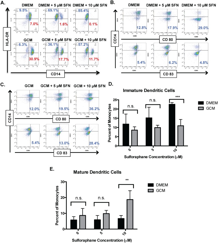
Fig 6. SFN promotes dendritic cell development from monocytes in both fresh media and GCM.
All experiments performed under hypoxic (1% O2) conditions. A) Representative dot plots showing CD14 and HLA-DR expression in CD14+ monocytes cultured in fresh media (DMEM) or BT116 GCM +/- SFN. Note that in addition to reductions in CD14+/HLA-DR- mMDSC’s (percent shown in red), there is a SFN-dependent increase in CD14- / HLA-DR+ cells suggestive of dendritic cells (percent shown in blue) in both fresh media and to a lesser extent in GCM. B) Representative dot plots showing CD14, HLA-DR, CD80, and CD83 expression in CD14+ monocytes cultured in DMEM +/- SFN. C) Representative dot plots showing CD14, HLA-DR, CD80, and CD83 epxression in CD14+ monocytes cultured in BT116 GCM +/- SFN. D) Bar graphs showing mean frequency of immature dendritic cells (CD14-/HLA-DR+/CD80+/CD83-) after culturing CD14+ monocytes from three donors in fresh media (DMEM) or BT116 GCM +/- SFN. Note that there is a SFN dose-dependent increase in immature dendritic cells with fresh media. E) Bar graphs showing mean frequency of mature dendritic cells (CD14-/HLA-DR+/CD80+/CD83+) after culturing CD14+ monocytes from three donors in fresh media (DMEM) or BT116 GCM +/- SFN. Note that there is a SFN dose-dependent increase in mature dendritic cells with GCM. n.s. = not significant, ** = P<0.01, *** = P<0.001.

