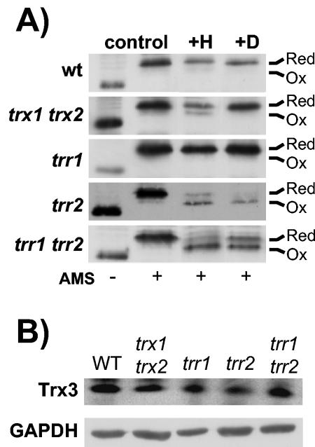
FIG. 6.
Redox state of Trx3 in thioredoxin mutants. (A) The indicated strains were grown to exponential phase in SD medium (control) and treated with 2 mM H2O2 for 1 h (+H) or 2 mM diamide for 1 h (+D). Proteins were precipitated with TCA, and free thiols were modified by reaction with AMS. Samples were separated using SDS-18% PAGE, and Trx3 was detected by Western blot analysis. Oxidized and reduced proteins are indicated. Trx3 contains two redox active Cys residues, as well as two additional Cys residues, and fully oxidized Trx3 was never detected. (B) Western blot analysis of Trx3 protein levels. Trx3 protein concentrations were measured in wild-type and trx1 trx2, trr1, trr2, and trr1 trr2 mutant strains by Western blot analysis with antibodies specific for c-Myc (Trx3) and glyceraldehyde 3-phosphate dehydrogenase (GAPDH).

