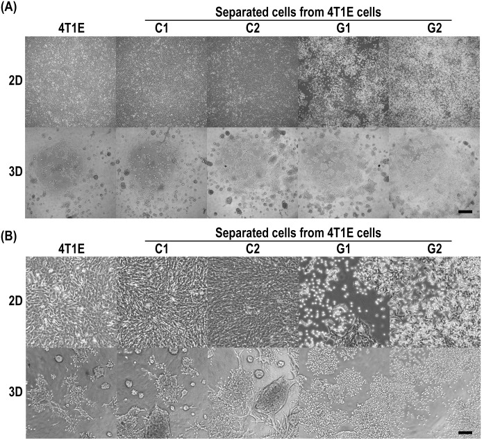
Fig 2. Microscopic images of 4T1E cells, and colony- and granular-type cells separated from 4T1E cells.
The separated cells were established as colony- (C1 and C2) and granular-type (G1 and G2) cells by optical separation based on morphology in PD-gelatin hydrogels. 2D, cell culture on a culture dish; 3D, cell culture in the PD-gelatin hydrogels. The images were taken after 3 days of culture under 2D and 3D conditions. (A) Raw images. The scale bar indicates 500 μm. (B) Digitally magnified images. The scale bar indicates 125 μm.

