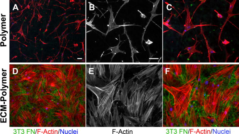
Figure 5. Adhesion and spreading of MSCs on a hybrid ECM-polymer scaffold.

Human MSCs were plated on a hybrid ECM – polymer scaffold or on a polymer scaffold without ECM. Cells were allowed to spread for 24 h, then cell shapes were visualized by staining actin filaments with rhodamine-phalloidin (red) and DAPI (blue). Samples were also stained with anti-fibronectin antiserum to show the NIH 3T3 matrix within the scaffold. B, C, E, F are higher magnification images of the samples in A and D. Scale bars = 50 μm.
