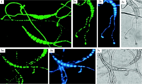
FIG. 7.
Nuclear localization of CHAP1-GFP fusion protein upon activation. 1. Conidia germinating on glass surface prior to hydrogen peroxide treatment; 2 and 3, germinated conidia treated with 50 mM hydrogen peroxide after 15 min of incubation. 2a and 3a, visualization of GFP-CHAP1 fusion protein by GFP fluorescence; 2b and 3b, DAPI staining of fungal nuclei; 2c and 3c, light microscope image. GFP fluorescence was visualized with an MRC-1024 laser confocal scanning microscope (Bio-Rad, Hempstead, United Kingdom) with a Nikon Plan Fluor 40/1.30 objective (Tokyo, Japan). Image processing was performed with Confocal Assistant 4.02 software (Bio-Rad, Hampstead, United Kingdom). DAPI fluorescence was visualized with a Zeiss Axioskop fluorescence microscope.

