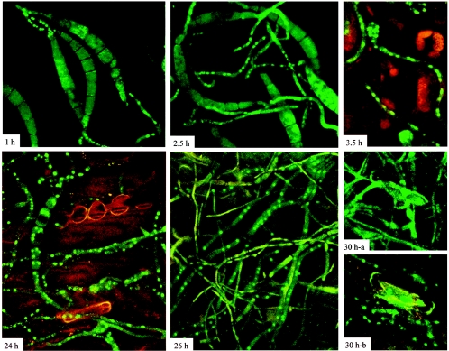
FIG. 9.
Subcellular localization of CHAP1 during plant infection. Numbers indicate time after inoculation: 1 h, conidia start germination, CHAP1 is activated in emerging germ tubes, and germ tubes elongate (2.5 h) and form appressoria (3.5 h); 24 h and 26 h, extensive mycelial network interlaces plant tissue, necrotic lesion is formed at the site of inoculation, fluorescence is localized in the nucleus in most of the hyphae; 30 h-a, hyphae found in the outermost layers of the infected leaf: GFP fluorescence is distributed in the cytosol; 30 h-b, hyphae found inside the plant tissue: in these hyphae CHAP1 is still activated. Confocal image acquisition was done as described for Fig. 7; the autofluorescence of the leaf, contributed primarily by chlorophyll, was detected with the Cy5-optimized channel.

