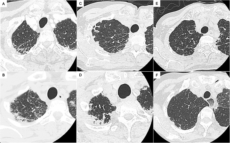
Fig 2. Representative chest computed tomography images of radiologic pleuroparenchymal fibroelastosis (PPFE)-like lesion.
Images of the right lung apex in patient 2 (A), patient 9 (B), patient 11 (C), patient 16 (D), patient 17 (E), and patient 20 (F) are shown. The images (E) and (F) reveal minimal cases of PPFE-like lesion we defined. Each patient number is listed in S1 Table.

