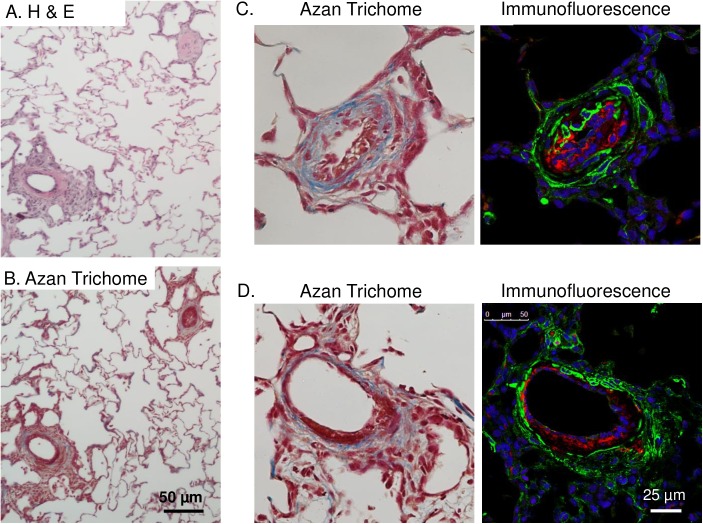
Fig 4. Hypoxia/SU5416 treatment causes both neointimal proliferation of endothelial cells and early plexiform lesions with collagen deposition.
All images are from hypoxia/SU5416-treated rats. (A-B) Representative H&E- and Azan trichrome-stained lung sections. (C-D) Higher magnification images of AZAN-stained and fluorescently-labeled arteries showing medial fibrosis and neointimal proliferation of endothelial cells and an early plexiform lesion. AZAN trichrome shows cell nuclei (dark red), collagen (blue) and orange-red in cytoplasm. Fluorescence labeling shows smooth muscle α-actin (green), Von Willebrand factor (red), and sytox (blue).

