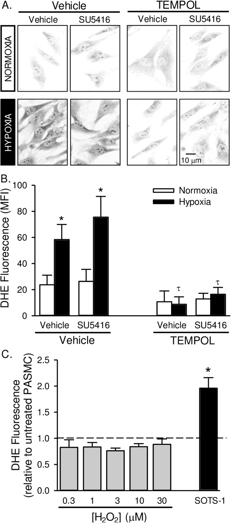
Fig 7. Superoxide levels are increased in pulmonary artery smooth muscle cells from pulmonary hypertensive rats.

Representative images (A) and summary data (B) showing background-subtracted mean fluorescence intensity (MFI) of dihydroethidium (DHE) in pulmonary arterial smooth muscle cells from rats treated with SU5416, TEMPOL or vehicle and exposed to normoxia or hypoxia. Fluorescence images were digitally inverted to provide improved signal contrast. Values are means ± SE; n = 5 animals per group; *P < 0.05 vs. corresponding normoxia group; τ p < 0.05 vs. corresponding TEMPOL-vehicle group; analyzed by multiple two-way ANOVA and individual groups compared with the Student-Newman-Keuls test. C) DHE fluorescence in PASMC following pre-incubation with increasing doses of H2O2 (0.3–30 μM) or SOTS-1 (10 μM). Dotted line represents untreated cells. Values are means ± SE; n = 5 animals per group; *P < 0.05 vs. untreated cells; analyzed by one-way ANOVA.
