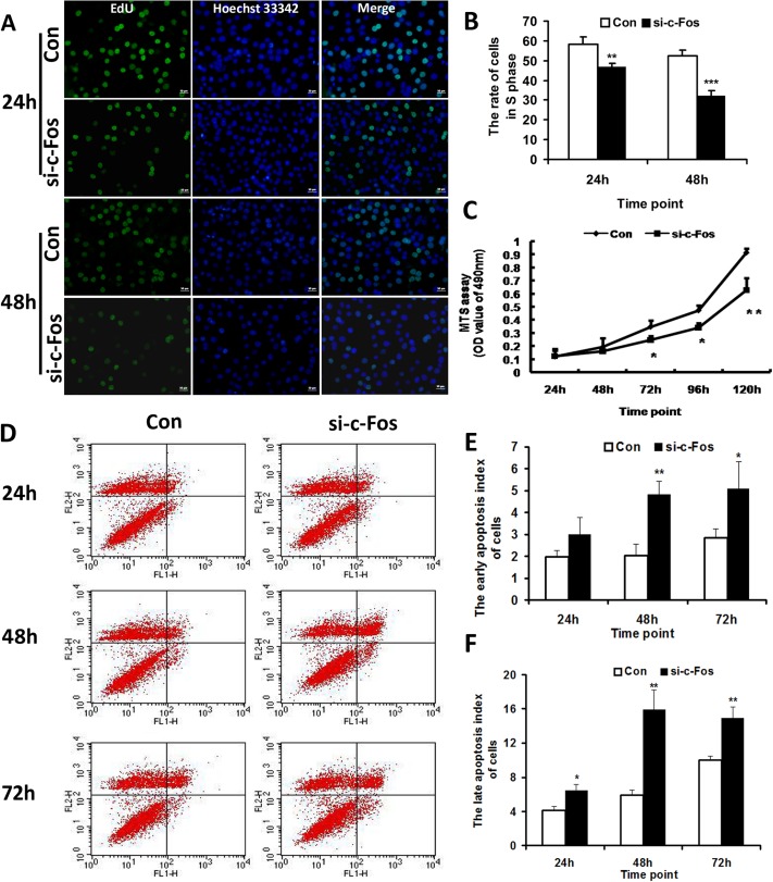
Fig 3. Knockdown of c-Fos inhibited the proliferation and promoted the apoptosis of MG63 cells.
A. Images of EdU DNA proliferation in vitro detection showing the cells in S phase treated with c-Fos siRNA (si-c-Fos) or the negative control (Con). bar = 50 μm. B. The rate of cells in S phase. Data represented means ± SD for three independent experiments of EdU DNA proliferation in vitro detection. C. MTS assay demonstrated that silencing of c-Fos inhibited the cell proliferation capability of MG63 cells on the indicated time points after transfection with c-Fos siRNA (si-c-Fos). D. Flow cytometer analysis showing that the apoptosis index of cells treated with c-Fos siRNA (si-c-Fos) was significantly increased compared with the control group. E. The early apoptosis index of cells. Data were from three independent experiments of flow cytometer analysis. F. The late apoptosis index of cells. Data were from three independent experiments of flow cytometer analysis. *p < 0.05, **p < 0.01, ***p < 0.001 versus negative control (Con).

