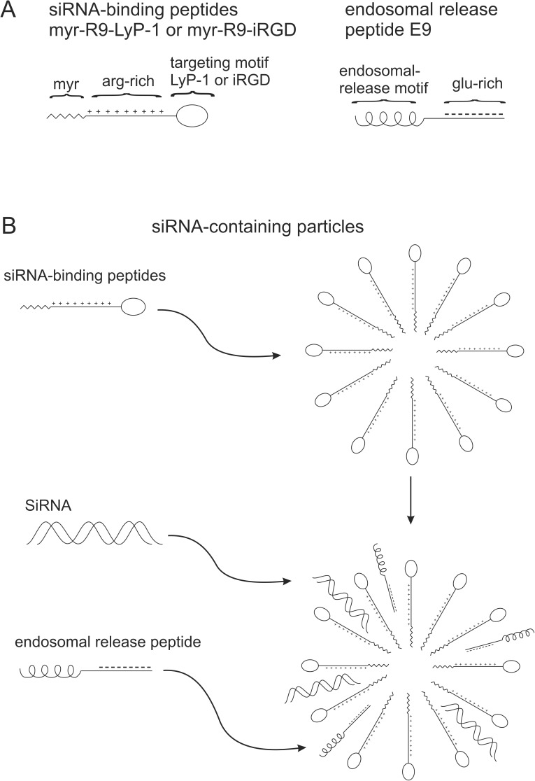
Fig 1. Diagrams of various peptides used in the study and the putative structure of complexes formed between the peptides and siRNA.
In Fig 1A, the domain structure of the peptides is shown. The siRNA binding peptides have a myristoylated amino-terminus, followed by an arginine-rich region (R9), and a cell targeting motif (LyP-1 or iRGD). The endosomal release peptide possesses an endosomal release motif flanked by a glutamic acid-rich region. In Fig 1B, the formation of nanoparticles by the siRNA-binding peptide is shown, and the incorporation of siRNA and the endosomal release peptide into the particles after their addition.

