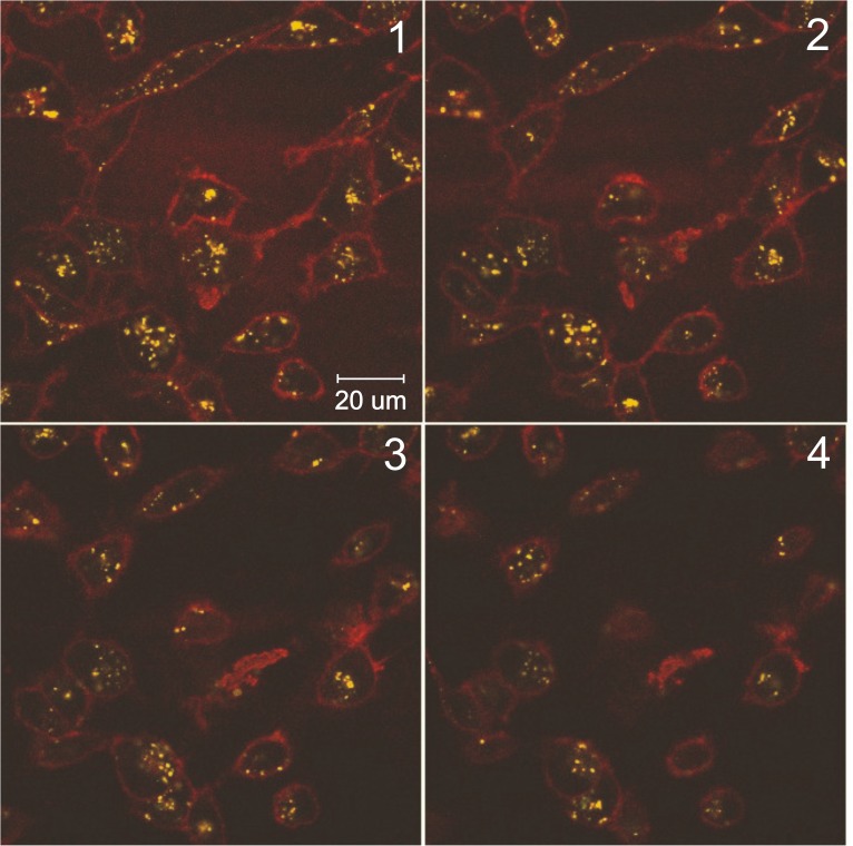
Fig 6. Confocal microscopy of MDA-MB-231 cells incubated with myr-R9-LyP-1/siRNA complexes.
Peptide/siRNA complexes were formed as described in the material and methods using fluorescently-labelled siRNA-binding peptide. The complexes were incubated on MDA-MB-231 cells for 18 hours, and the cells were then live-mounted onto glass slides. The slides were viewed by confocal microscopy, and serial sections through one field of cells are shown (panels 1–4). The fluorescently-labeled siRNA-binding peptide is shown in yellow, and the plasma membrane is shown in red (Cell Mask plasma membrane stain). The results presented are representative of three independent experiments.

