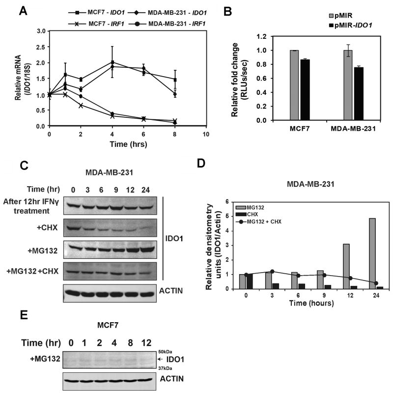
Figure 5.
Post-transcriptional regulation of IDO1 expression in BC cells. A, Analysis of IDO1 mRNA stability in MDA-MB-231 and MCF7 cells after both cell lines were stimulated with IFNγ (100ng/mL) for 4 hrs followed by treatments with actinomycin-D (1 μg/mL), a transcription inhibitor. B, Analysis of 3′-UTR based post-transcriptional regulation of IDO1 expression with a reporter in which the 3-UTR region of IDO1 was cloned into pmirGLO 3′-UTR reporter plasmid. C, Immunoblot analysis of the protein stability of IFNγ-induced IDO1 after treatment with CHX (50 μg/mL) and MG132 (5 μM) in MDA-MB-231 cells. D, Densitometric analysis of panel C for IDO1 expression in MDA-MB-231 cells. E, Immunoblot analysis of the protein stability of IFNγ-induced IDO1 after treatment with MG132 (5μM) in MCF7 cells. Data in A and B are shown by mean ± SEM from three independent measurements. Results in C, D and E are representative of at least two independent experiments.

