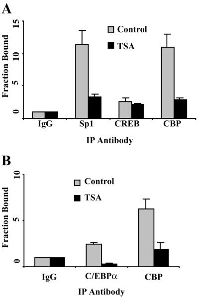
FIG. 7.
The Sp1 site within the bcl-2 P1 promoter and the Cdx site within the bcl-2 P2 promoter are involved in TSA-induced bcl-2 transcriptional repression. (A) Quantitative ChIP assays of the binding of Sp1, CREB, and CBP to the bcl-2 P1 promoter. DHL-4 cells were treated with 500 ng of TSA/ml for 18 h. ChIP assays were performed as described in the legend to Fig. 6B, except that antibodies specific to Sp1, CREB, and CBP were used. The primers for the bcl-2 P1 promoter are shown in Fig. 4A. The fraction bound represents the fold increase of the amount of the bcl-2 P1 promoter in the specific antibody immunoprecipitated (IP) DNA compared to the level of nonspecific IgG antibody-immunoprecipitated DNA. (B) Quantitative ChIP assays of the binding of C/EBPα and CBP to the bcl-2 P2 promoter. The assays were performed as described for panel A with antibodies specific to C/EBPα and CBP. The primers for the bcl-2 P2 promoter are shown in Fig. 4A.

