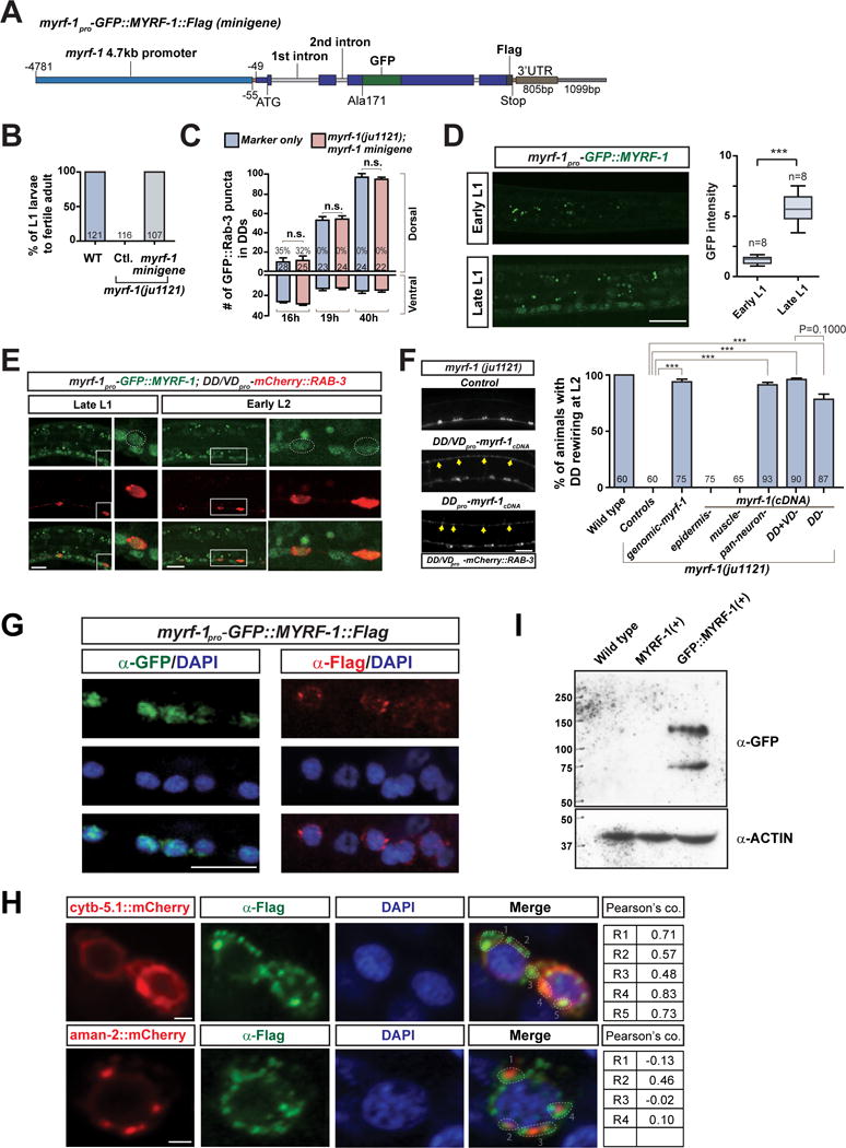
Figure 3. Dual localization of MYRF-1 in cytoplasm and nucleus.

A. Illustration of myrf-1pro-GFP∷MYRF-1∷Flag minigene transgene.
B. Percentage of myrf-1(ju1121) mutants carrying myrf-1 minigene (ybqIs13) (picked at L1) that developed into fertile adults.
C. Number of synapses is quantified for myrf-1(ju1121) carrying myrf-1 minigene transgene (ybqIs13), labeled by flp-13pro-GFP∷Rab-3 (ybqIs47), and shown as mean ± SEM; t-test (n.s.); the number of animals analyzed is shown on each bar.
D. GFP signal from myrf-1 minigene (ybqIs13) increased at late L1. Arrows, ventral cord neurons with GFP signal. GFP signal in soma normalized by background signal is shown as mean ± SEM (box bars); t-test (***P<0.001); Line bars, min/max values; n, number of images analyzed.
E. Co-localization of native GFP signal from GFP∷MYRF-1 (ybqEx164) and unc-25pro-mCherry∷RAB-3 (juIs236).
F. Tissue specific expression of myrf-1 cDNA in myrf-1(ju1121) mutants. Genomic myrf-1 (ybqEx55), 11.5 kb amplified from genomic DNA; DD flp-13 (ybqEx94); DD/VD unc-25 (ybqEx85); epidermis dpy-7 (ybqEx16); body wall muscle myo-3 (ybqEx93); pan-neuron rgef-1 (ybqEx86). Percentage of animals with rewired DDs is shown as mean ± SEM; t-test (***P < 0.001); the number of animals analyzed is shown on each bar. Arrows, dorsal DD synapses.
G. Immunostaining of myrf-1 minigene transgene (ybqIs13) using anti-GFP and anti-Flag. DAPI, a DNA-binding dye. Scale, 10 μm.
H. Immunostaining using anti-Flag on dual-transgene animals with myrf-1 minigene (ybqIs13) and pan-neuronally (rgef-1pro) expressed ER marker cytb-5.1∷mCherry transgenes (ybqEx595); or Golgi marker aman-2∷mCherry (ybqEx597). Red and green signals in each ROIs are test for co-localization; R, Pearson’s coefficiency. Scale, 1 μm.
I. Protein extracts from N2, myrf-1(+), and GFP-myrf-1(+) transgene animals (ybqEx55, ybqIs13) are analyzed by Western Blot. Two bands (140 kDa, 75 kDa) are detected by anti-GFP. (See also Figure S3, S4)
