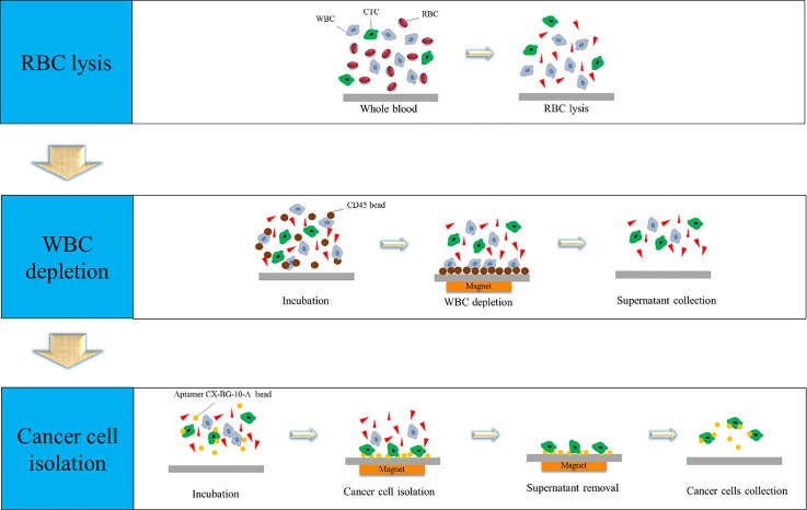
FIG. 1.
Schematic diagram of the CTC isolation process including (1) RBC lysis, (2) WBC depletion, and (3) CTC capture. RBC lysis buffer was first used to lyse RBCs. Then, anti-CD45 magnetic beads were used to deplete WBCs, and the supernatant containing the cancer cells was collected. Finally, aptamer-coated magnetic beads were used to isolate the CTCs in the supernatant from the previous step.

