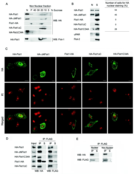
FIG. 2.
Subcellular localization and interaction with PTOV1 of HA-tagged forms of flotillin-1. (A) Association of epitope-tagged flotillin-1 variants with lipid rafts. Nuclear and nonnuclear fractions were obtained from PC-3 cells transfected with the indicated variants. Nonnuclear fractions were further fractionated by sucrose density gradient (Fig. 1). The fractions (5 to 40% of sucrose), the pellet of the gradient (P), and the total crude nuclear extract (N) were analyzed by Western blotting (WB) with anti-HA. The same membranes were reanalyzed for endogenous flotillin-1 (Flot1) partitioning and gave results similar to those of Fig. 1C (endogenous flotillin-1 is shown only for the HA-Flot1 transfectant). (B) Nuclear localization of epitope-tagged variants of flotillin-1. (Left panel) Cells were transfected with the indicated plasmids, and 10% of the crude nuclear extract (N) and of nonnuclear fractions (S) were analyzed by Western blotting with anti-HA antibody. The same membranes were reanalyzed with antibodies to uPAR and to flotillin-2 and found to be negative for plasma membrane protein contamination of nuclear extracts(shown are the HA-Flot1 transfectants). (Right panel) In parallel transfections, the percentage of cells with nuclear HA was assessed for each construction, as recorded by immunocytochemistry. At least 200 cells were counted in each case in two independent experiments. (C) Representative confocal immunofluorescence images of transfected flotillin-1 variants stained with anti-HA (top) and with propidium iodide (PI) (bottom). Control reactions eliminating the primary antibody gave negative staining. Scale bars, 10 μm. (D) Coimmunoprecipitation of FLAG-PTOV1 and flotillin-1 variants. Cells were cotransfected with pFLAG-PTOV1 and HA-tagged flotillin-1 variants, lysates were immunoprecipitated with anti-FLAG antibody, and total cell extracts (10% [Input]), IP, nonprecipitated material (10% [S]), or precipitated proteins from a control antibody (data not shown) were detected by Western blotting with anti-HA and anti-FLAG antibodies. (E) Coimmunoprecipitation of FLAG-PTOV1 and HA-Flot1 in nuclear or nonnuclear fractions. Lysates from cells cotransfected with pFLAG-PTOV1 and pHA-Flot1 were separated into nuclear and nonnuclear fractions and immunoprecipitated with anti-FLAG, and coimmunoprecipitates were detected by Western blotting with anti-HA and anti-FLAG antibodies.

