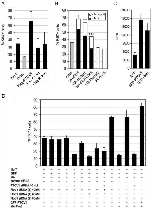
FIG. 6.
PTOV1 and flotillin-1 are required for normal cell proliferation. The effect of overexpression of exogenous PTOV1 or flotillin-1 or the depletion of the corresponding endogenous proteins on the proliferative state of PC-3 cells was assessed by double fluorescence for GFP, anti-FLAG or anti-HA, and anti-Ki67. At least 200 cells were scored for each condition. The results shown are the means from three independent experiments for each condition. (A) Mitogenic effects of full-length PTOV1 and the two separate PTOV domains of PTOV1. FLAG constructs were transfected, and cells were analyzed 48 h after transfection by double fluorescence staining with anti-Ki67 and anti-FLAG antibodies. For mock transfections with empty FLAG vector and untransfected cells (No T), only the Ki67 staining was scored. (B) Mitogenic activity of flotillin-1 variants. Forty eight hours after transfection of the indicated plasmids, cells were analyzed by double fluorescence for Ki67 and HA reactivities to determine their proliferative capacities. Nuclear (N) or nonnuclear (Non N) staining of the HA reactivity was also scored to determine the capacity for each variant to enter the nucleus. Significance for Ki67 values relative to mock controls is as follows: *, P = 2.97 × 10⁻⁶; **, P = 0.0001; ***, P = 0.19. (C) Overexpression of GFP-flotillin-1 induces cell proliferation. PC-3 cells were transfected with pEGFP, pGFP-PTOV1, or pGFP-Flot1 and analyzed 48 h after transfection for [3H]thymidine incorporation. Induction of proliferation for each plasmid, corrected for transfection efficiencies, is expressed as counts per minute (CPM). (D) Effects of the depletion of PTOV1 or flotillin-1 on the proliferative status of PC-3 cells. Cells were transfected with pEGFP or pCMV-HA as controls, pGFP-PTOV1, or pHA-Flot1 and the indicated siRNAs and assessed by double fluorescence detection for Ki67 and GFP or HA. For cells transfected with pGFP-PTOV1 and pHA-Flot1, fluorescent staining for Ki67 was determined for GFP-positive cells.

