Figure 3.

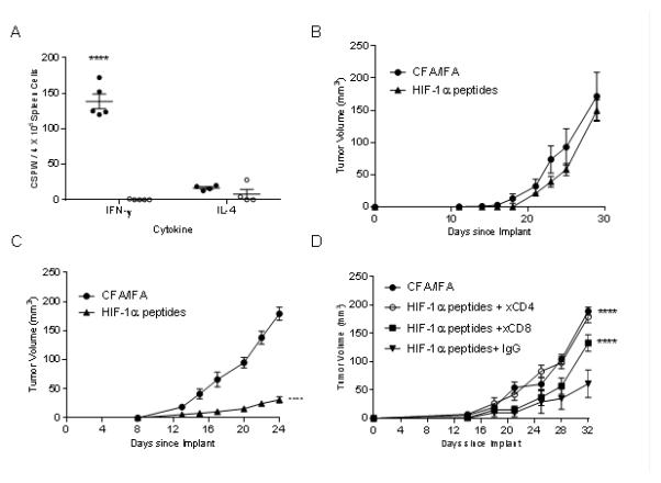
HIF-1α poly-epitope vaccines are significantly more effective in inhibiting growth of basal than luminal mammary tumors. (A) IFN-γ and IL-4 ELISPOT in splenocytes after completion of three immunizations. Antigens include a pool of the HIF-1α vaccinating peptides (●), HIV p52 (○) as a negative control and ConA (▲) as a positive control. The data are presented as corrected spots per well (CSPW). The horizontal bar indicates the mean CSPW ± SEM. n=5 mice/group; ***p<0.001 compared to HIV. Mean tumor volume (mm3 ± SEM) from mice injected with adjuvant alone (●) or HIF-1α poly-epitope vaccine (▲) in TgMMTV-neu mice (B) or C3(1)Tag mice (C), n=5 mice/group; ****p<0.0001. (D) Mean tumor volume (mm3 ± SEM) from C3(1)Tag mice injected with adjuvant alone (●), or HIF-1α poly-epitope vaccine treated with mouse IgG (●), anti-CD4 (○) or anti-CD8 (■) n=5 mice/group; ****p<0.0001 compared to HIF-1α poly-epitope vaccine+IgG.
