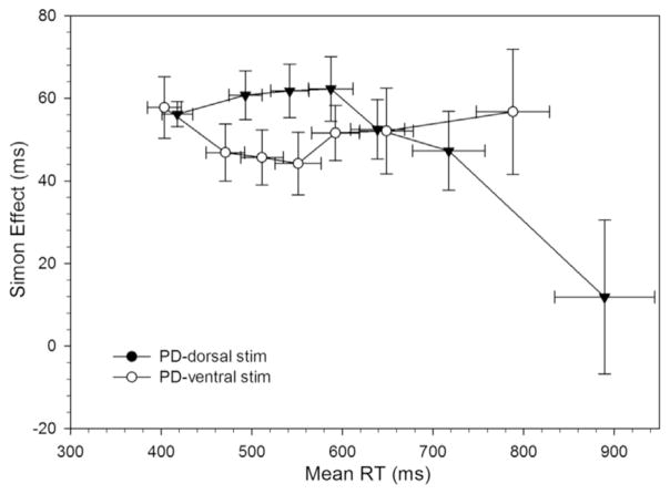
Fig. 10.
RT delta plots for Parkinson’s disease (PD) participants on dorsal and ventral STN stimulation. The reduced proficiency of suppressing interference on ventral stimulation is significantly improved in with dorsal stimulation (i.e., a larger negative-going delta slope at the slow end of the distribution).

