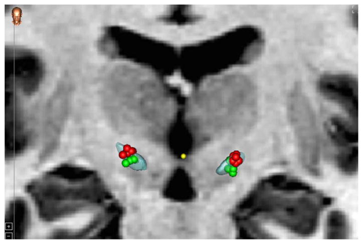
Fig. 4.
Distribution of the electrode contacts used, and projected volume of tissue activation with .4 mA, 130 Hz and 60 μs, for dorsal (red) and ventral (green) stimulation of the STN. (For interpretation of the references to color in this figure legend, the reader is referred to the web version of this article.)

