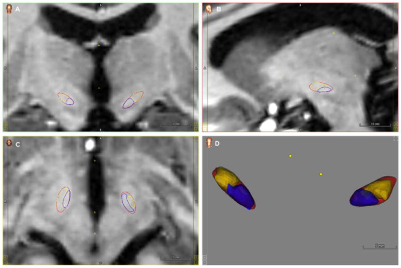
Fig. 5.
Subdivision of the STN in dorsal (yellow) and ventral (purple) to determine contact localization at different planes. Visualizations were projected on Coronal (A), Sagittal (B) and Axial plane (C) and a 3D volume (D). (For interpretation of the references to color in this figure legend, the reader is referred to the web version of this article.)

