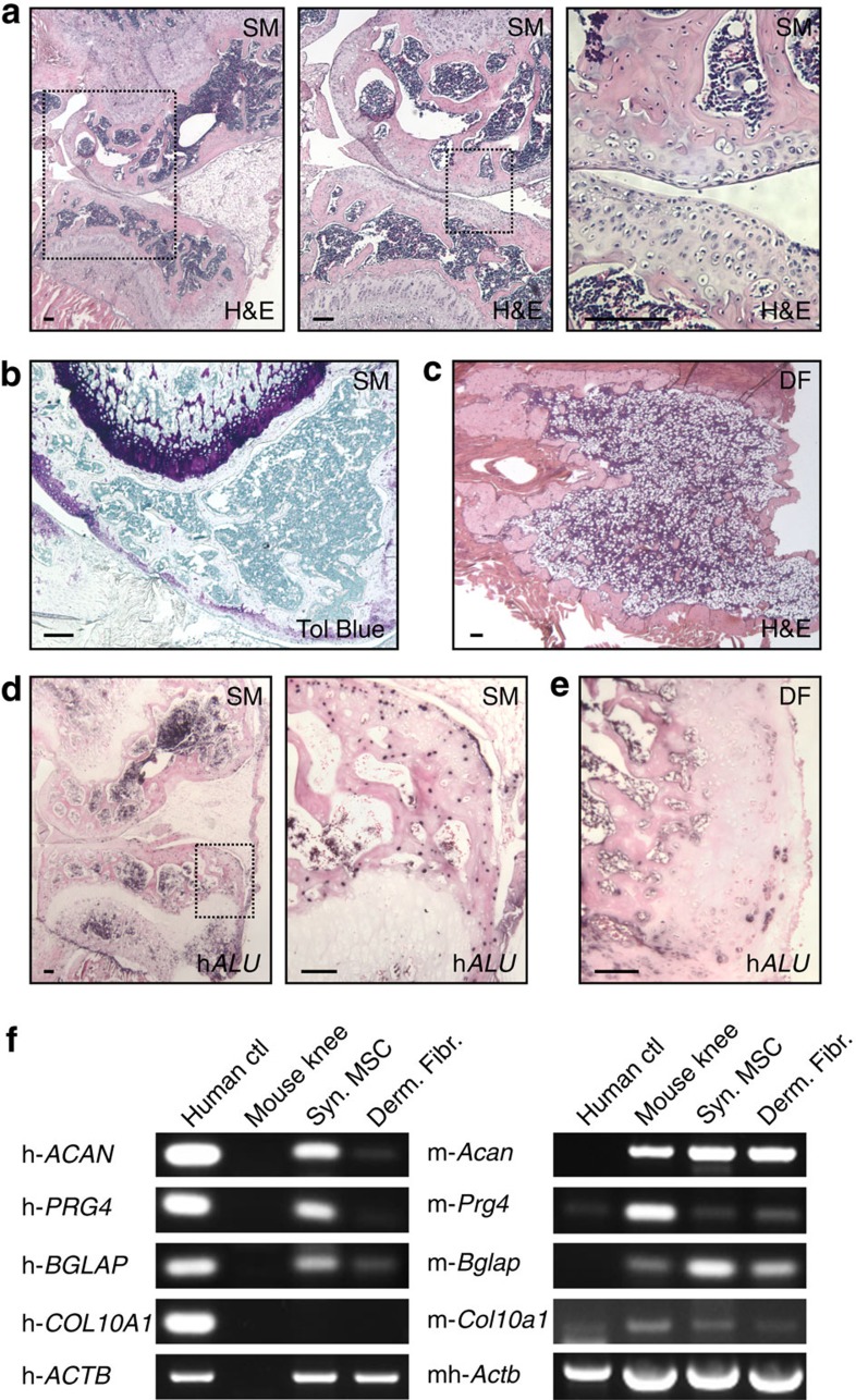
Figure 8. Joint morphogenetic properties of adult human synovial MSCs.
(a,b,d,f) Human synovial MSCs (n=1) transduced with Ad-Bmp7 and injected into nude mouse thigh formed a joint-like structure upon explant 4 weeks later, while (c,e,f) dermal fibroblasts (n=1) transduced with Ad-Bmp7 gave rise to an ossicle. (d,e) ISH for human-specific ALU genomic repeats. Black dots indicate human nuclei. Scale bars, 100 μm. Boxed areas on left shown at higher magnification on right. SM: synovial MSCs; DF: dermal fibroblasts; H&E: haematoxylin and eosin; Tol blue: toluidine blue; hALU: human ALU repeats. (f) RT–PCR analysis using species-specific primers. Human positive controls (human ctl) were freshly isolated chondrocytes from articular cartilage for ACAN (aggrecan) and PRG4 (lubricin), MSCs treated with vitamin D3 for BGLAP (osteocalcin), and bone marrow MSCs treated with TGFβ1 in pellet culture for COL10A1 (collagen type X); a mouse knee joint was used as mouse ctl for all genes. h: primer pair specific for human; m: primer pair specific for mouse; mh: primer pair detects both mouse and human. Samples were equalized for h-ACTB (h-β-actin; human ctl used for h-ACAN and h-PRG4 is shown), or for mh-Actb in the case of the mouse ctl. See also Supplementary Fig. 9.

