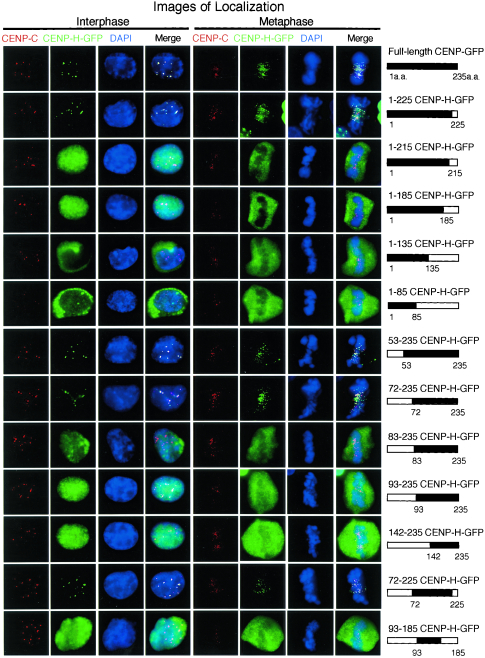
FIG. 2.
Identification of the minimal region required for CENP-H function. A schematic representation of CENP-H-GFP constructs and summary of the complementation and localization analysis are shown. To examine localization of CENP-H derivatives, an anti-CENP-C antibody was used to stain centromeres. Typical images for immunofluorescence are shown. Antibody signals were detected with Cy3-conjugated secondary antibodies (red), and GFP signals appear green. DNA was counterstained with DAPI (blue).

