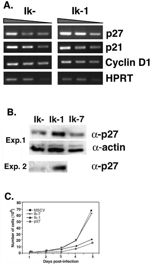
FIG. 5.
Ik-1-transduced JE131 cells display an increase in p27kip1 expression and can be slowed in their growth by retroviral transduction of p27kip1. JE131 cells with and without Ik-1 activity were analyzed for RNA and protein expression of p27kip1. (A) At 24 h after infection with the MSCV-IRES-GFP (Ik−) or MSCV-(Ik-1)-IRES-GFP (Ik+) retrovirus, successfully transduced JE131 cells were purified and cDNA was prepared. RT-PCR was performed by using a range of cDNA amounts. Shown here are the results of twofold dilutions (from 1:256 to 1:1024 for each sample). (B) At 24 h after infection with MSCV-(Ik-1)-IRES-GFP (Ik-1), MSCV-(Ik-7)-IRES-GFP (Ik-7), or MSCV-IRES-GFP (Ik−) retrovirus, successfully transduced JE131 cells were purified, and protein extracts were prepared. A total of 10 μg of protein was subjected to SDS-PAGE. Blots were probed with antibodies against p27kip1 or actin (as a loading control). Results from two representative experiments are shown. (C) JE131 cells were infected with the MSCV-IRES-H-2Kk (Ik−), MSCV-(Ik-7)-IRES-H-2Kk (Ik-7), MSCV-(Ik-1)-IRES-H-2Kk (Ik-1), or MSCV-(p27kip1)-IRES-H-2Kk (p27) retrovirus. Successfully transduced cells were purified and plated at 106 cells/well in a 24-well plate. Counts of viable cells were performed every 24 h. This figure was generated by using CellQuest, Microsoft Excel, and Adobe Photoshop software.

