FIG. 4.
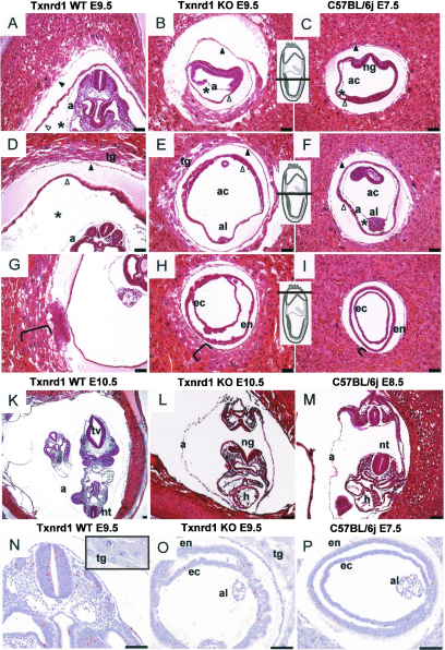
Histological analysis of paraffin-embedded Txnrd1-deficient and wild-type embryos. (A to I) Serial sections of hemizygous Txnrd1 (A, D, and G) and homozygous Txnrd1 KO (B, E, and H) siblings at E9.5 as well as corresponding wild-type embryos at E7.5 (C, F, and I). Inserts indicate section levels. The brackets in panels G, H, and I indicate the layering of trophoblast giant cells. Whereas the homozygous KO embryo had retarded growth and resembled E7.5 wild-type embryos, extraembryonic tissues seemed unaffected by the Txnrd1 deficiency. Asterisks indicate the exocoelomic cavity, black arrowheads mark the Reichert's membrane, and white arrowheads point to the yolk sac. (K to L) Cross sections of hemizygous Txnrd1 (K) and homozygous Txnrd1 KO (L) siblings at E10.5 compared to a wild-type embryo at E8.5 (M). Note that the open neuralgroove of the homozygous KO embryo (L) is opposed to the caudal neuropore, indicating that turning had not occurred. (N to P) Mitotically active cells were detected by phospho-histone H3 (PH3) immunostaining (brown color). Hemizygous embryos at E9.5 (N) displayed significantly more mitotically active cells than Txnrd1 KO embryos of the same age (O), whereas the number of PH3-positive cells at E9.5 in Txnrd1 KO embryos was similar to that in WT embryos at E7.5 (P). Sections A to M were stained with hematoxylin and eosin, and sections N to P were counterstained with hematoxylin after immunodetection. Bars = 1,000 μm. Abbreviations: a, amnion; ac, amniotic cavity; al, allantois; ec, ectoderm; en, endoderm; h, embryonic heart; ng, neural groove; nt, neural tube; tg, trophoblast giant cells; tv, telencephalic vesicle.

