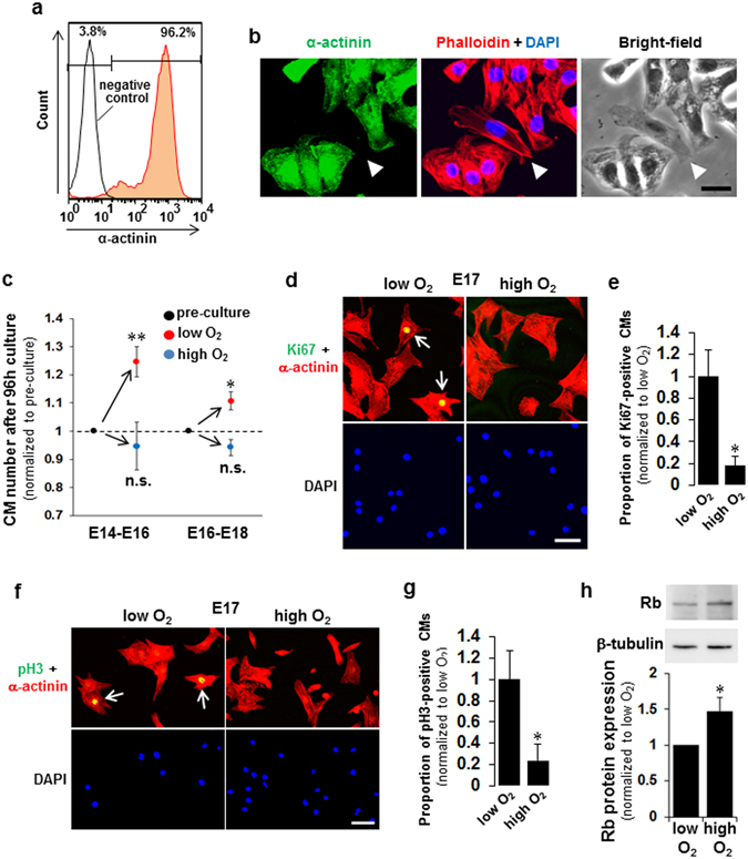
Figure 1.

Exposure of fetal cardiomyocytes (fCMs) to O2 inhibits proliferation and cell cycle activity. (a) FACS analyses of E16 fCMs stained for α-actinin indicate > 95% purity when isolated and cultured under low O2 conditions. (b) Immunofluorescence for α-actinin observed in phalloidin, DAPI, and bright-field images of E16 fCMs. The arrowhead indicates one of the few contaminating fibroblasts, which have a distinct phase appearance and are completely negative for α-actinin. (c) The fCMs (E14–E16 or E16–E18) isolated under low O2 conditions were cultured under low or high O2 conditions for 96 h. At the start and end of the culture, total cell numbers were counted, and the proportion of CMs and non-CMs were determined by FACS to obtain the absolute number of each cell type. n = 3–5 independent experiments. * P < 0.05 and ** P < 0.01 compared to pre-culture levels. n.s. = not significant. (d,f) Immunofluorescence for Ki67 (d) and pH3 (f) observed in α-actinin and DAPI of low O2-isolated fCMs cultured under low or high O2 conditions. Arrows denote Ki67- (d) and pH3- (f) positive fCMs. (e,g) Proportions of Ki67- (e) and pH3- (g) positive CMs in (d) and (f), respectively. n = 5 independent experiments. * P < 0.05. (h) Rb protein expressions in low O2-isolated fCMs (E16–E17) cultured under low or high O2 conditions. β-tubulin was used as a loading control. n = 5 independent experiments. * P < 0.05. Error bars = SEM. Scale bars = 20 µm in (b) and 50 µm in (d,f).
