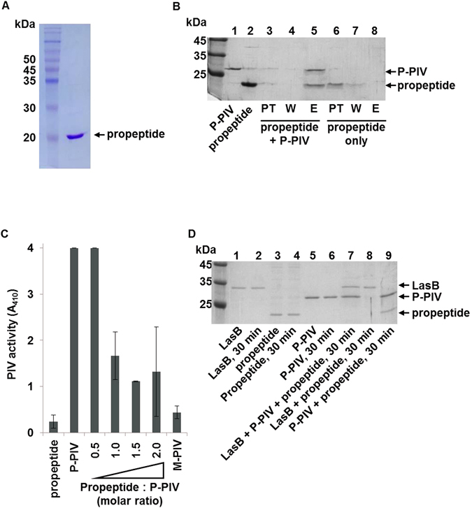
Figure 5.

Binding of propeptide to PIV and inhibition of PIV activity. The propeptide of PIV was purified from E. coli (A). (B) 1 μg of the purified propeptide was mixed with 1 μg of P-PIV in 25 μl reaction volume and incubated at room temperature for 10 min. The mixture was loaded to Ni-NTA agarose affinity column, washed twice with 5 mM imidazole-containing washing buffer, and eluted by 200 mM imidazole-containing elution buffer (lanes 3–5). Same amount of the purified propeptide was applied to Ni-NTA agarose affinity column in the same manner (lanes 6–8). PT, W, and E indicate fractions of pass through, wash, and elute, respectively. The purified propeptide of PIV and P-PIV were loaded as control (lane 1, 2). This gel picture was cropped and the full-length gel picture is presented in supplementary data (Fig. S4C). (C) The purified propeptide was mixed with 100 ng of P-PIV at the indicated molar ratio and incubated in room temperature for 10 min. The PIV activity was then measured by chromogenic substrate. As controls, 160 ng of propeptide, 100 ng of P-PIV, or 100 ng of M-PIV were separately measured for the PIV activity for comparison. (D) 500 ng of the purified LasB, 1 μg of propeptide, and 1 μg of P-PIV were mixed in various combination and incubated at room temperature for 30 min. The proteins were analyzed in 12% SDS-PAGE and visualized by Coomassie staining. To know stability, each protein was directly applied to SDS-PAGE without incubation (lanes 1, 3, 5). This gel picture was cropped and the full-length gel picture is presented in supplementary data (Fig. S4D).
