
Figure 1.
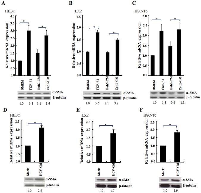
Conditioned medium from the culture of HCV replicon cells or HCV-infected cells increased procollagen I transcripts and α-SMA protein expression in HSC. (A–F) The HSCs of HHSC (panels A and D), LX2 (panels B and E) and HSC-T6 (panels C and F) were incubated with the conditioned medium (CM) derived from the 72 h culture of Huh7 and Con1 replicon cells (panels A,B and C), or the Huh7 cells infected with HCVcc (JFH1, MOI = 3; panels D,E and F). The level of procollagen I gene expression was quantified by real-time RT-PCR and the expression of β-tubulin was used as a control for normalization. Histograms were used to show the relative expression levels of procollagen I mRNA among different treatments. The cells cultured in DMEM without any treatment were arbitrarily denoted as 1. The data represent the mean ± S.D. (n = 3; *p < 0.05). In parallel, the cell lysates were harvested and Western blot analysis was performed using the anti α-SMA antibody. The ratios for the relative band intensities of α-SMA normalized by the expression of β-tubulin are shown at the bottom. HSCs cultured in the DMEM or DMEM supplemented with TGFβ1 (10 ng/ml) were used as the negative and positive control, respectively. The full Western blot and the corresponding positions of the molecular weight protein markers are presented in Supplementary Fig. S1.