Figure 5.
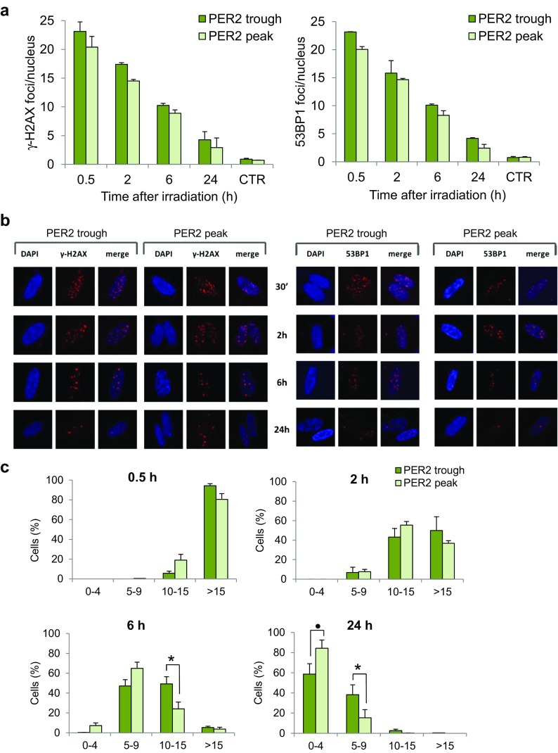
The kinetics of ionizing radiation-induced foci (IRIF) in serum-shocked γ-irradiated human CCD-34Lu fibroblasts. The mean number of γ-H2AX foci and 53BP1 foci (a) after irradiation administered at the trough and the peak of PER2 protein expression. b Representative immunofluorescence of γ-H2AX and 53BP1 foci in nuclei stained with DAPI at 0.5, 2, 6, and 24 h after irradiation. c Cells were categorized as having 0–4, 5–9, 10–15, and >15 of γ-H2AX and 53BP1 foci/nucleus. Data are means ± SD from independent experiments, each with at least 200 nuclei/time points, carried out in cells irradiated at the trough and the peak of PER2 protein expression (• p < 0.1, *p < 0.05; ANOVA test)

