FIG. 7.
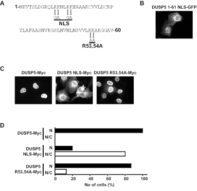
Identification of a functional NLS within the amino terminus of DUSP5. (A) Amino acid sequence of the first 61 residues of DUSP5. Mutations introduced into the putative arginine- and lysine-rich NLS and the conserved arginine residues of the KIM are indicated by arrows. (B) Mutation of the putative NLS within the DUSP5 1-61 NLS-GFP fusion prevents nuclear localization. (C) Myc-tagged wild-type DUSP5 is a nuclear protein, and mutation of the NLS but not the KIM prevents the nuclear accumulation of DUSP5. (D) The distribution of Myc-tagged proteins in at least 200 expressing cells for each transfection in panel C was recorded and is presented graphically as the percentage of cells in which fluorescence either was predominantly nuclear (N) or was found in both the nucleus and the cytoplasm (N/C). Experiments were performed at least three times, and representative images and cell counting results are shown.

