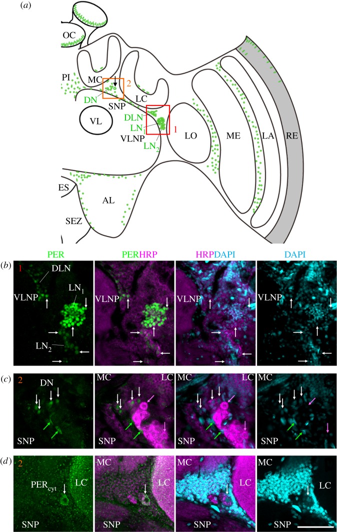
Figure 3.
amPER-immunoreactive neurons in the honeybee brain. (a) Schematic presentation of amPER-positive cells (green) in the right brain hemisphere. amPER-positive neurons were present in the dorsal and lateral central brain and named DN, DLN, LN1 and LN2 according to their location and the nomenclature in Drosophila melanogaster. The most conspicuous LN1 are shown together with the LN2 and DLN in (b) (red rectangle 1 in (a)) and in more detail in figure 4a,b and electronic supplementary material, figure S3). The DN and one amPER-positive cell with cytoplasmic staining (arrow in (a)) are shown in (c) and (d) (orange rectangle 2 in (a)). Note that (d) is located slightly posterior of (c). (b) amPER-positive neurons in the dorsolateral and lateral brain (DLN, LN1, LN2). White arrows point to selected neurons of all three groups. All PER cells were also marked by HRP and DAPI, although the staining was sometimes weak (overlay of two confocal stacks). (c) PER-positive cells in the dorsal brain between medial (MC) and lateral calyces (LC) and the superior neuropils (SNP). White arrows point to four PER-positive neurons, the cytoplasm of which is also labelled by HRP and the nuclei weakly by DAPI. Green arrows point to two PER-positive but HRP-negative glial cells, the nuclei of which are also labelled by DAPI. Magenta arrows point to two HRP-positive neurons that are PER-negative (one single confocal stack). (d) A large PER-positive neuron with cytoplasmic staining (PERcyt), which is located in the dorsal brain between medial (MC) and lateral calyces (LC) and the superior neuropils (SNP). The cytoplasm of this neuron is co-stained with HRP and its nucleus is weakly DAPI-positive (overlay of two confocal stacks). AL, antennal lobe; ES, esophageal foramen; LA, lamina; LC, lateral calyx; LO, lobula; MC, medial calyx; ME, medulla; OC, ocelli; PI, pars intercerebralis; RE, retina; SEZ, subesophageal zone; SNP, superior neuropils; VL, vertical lobe of the mushroom body; VLNP, ventrolateral neuropils. All photos in (b–d) were taken from foragers' brain. Scale bars, 30 µm. Pictures are taken with a 10× objective (numerical aperture: 0.3); distance of z-stacks 2.5; overlay of three stacks.

