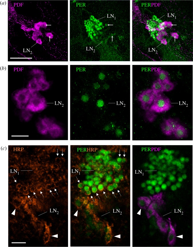
Figure 4.
A finer characterization of the LN2. (a) Lateral neurons LN1 and LN2 (overlay of 10 confocal stacks from a vibratome section, distance of z-stacks 1.0 µm). All LN2 are co-stained with both anti-PER and anti-PDF. White arrows point to selected LN2. Scale bar, 30 µm. (b) Magnification of the PDF-positive LN2 (single confocal stack from a wholemount brain; 10× objective; numerical aperture: 0.30). The brain was scanned in z steps of 2.5 µm, and the present image is at a depth of 85 µm from the anterior surface. PER is confined to the nuclei of the LN2. Scale bar, 10 µm. (c) Single confocal stack of a vibratome section (the same as in figure 3b) showing the LN1 and LN2 stained by anti-PER, anti-HRP and anti-PDF (same microscope settings as in (b)). HRP and PDF are shown in magenta and indicate the size of the neurons. Although HRP does not stain the cytoplasm membrane uniformly, it shows that the LN1 are much smaller than the LN2. All photos were taken from foragers' brains. Arrows point to single LN1 neurons the cytoplasm of which is clearly labelled by the neuronal marker HRP. Arrowheads point to single LN2 neurons the cytoplasm of which is HRP positive and additionally labelled by PDF. Scale bar, 10 µm.

