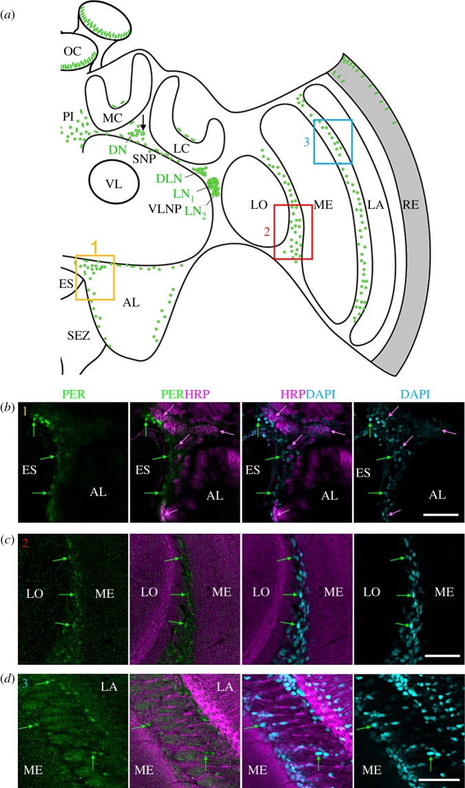
Figure 5.
Putative amPER-positive glia cells. (a) Schematic presentation of amPER-positive cells (green) in the right brain hemisphere. Putative amPER-positive glial cells were present throughout the cortex of the brain, dorsally of the calyces (MC and LC), at the border of the antennal lobe (AL) (yellow rectangle 1), between lobula (LO) and medulla (ME) (red rectangle 2) and between medulla (ME) and lamina (LA) (blue rectangle 3). (b) PER-positive glial cells around the esophageal foramen (ES) close to the antennal lobe (AL) (see inset 1 in (a)). Green arrows mark selected PER-positive nuclei that are also stained by DAPI but not by HRP. Selected HRP-positive neurons are marked by magenta arrows. The nuclei of the latter were also stained by DAPI, but clearly less intensively than the glial cells. (c) PER-positive glial cells between the lobula (LO) and medulla (ME) (see inset 2 in (a)). Labelling as in (a). (d) PER-positive glial cells between the medulla (ME) and lobula (LO) (see inset 3 in (a)). All photos in (b–d) were taken from foragers' brains. Please note that only subsets of glia cells are PER positive. Scale bars, 30 µm.

