
Figure 2. Western blot analysis of small-scale expression of FGFR2 and FGFR3 constructs using anti-MBP antibody.
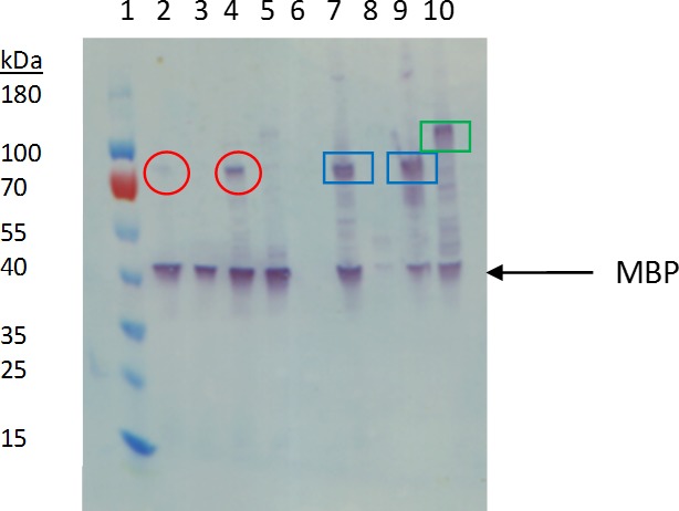
Lane 1: Ladder. Lane 2: FGFR2 31-406 from supernatant. Lane 3: FGFR2 370-651 from supernatant. Lane 4: FGFR3 143-405 from supernatant. Lane 5: FGFR 3: 365-771 from supernatant. Lane 6: Blank. Lane 7: FGFR2 31-406 from pellet. Lane 8: FGFR2 370-651 from pellet. Lane 9: FGFR3 143-405 from pellet. Lane 10: FGFR3 365-771 from pellet. Circled in red are bands consistent with FGFR2 and FGFR3 ECD + TM from supernatant. Boxed in blue are bands consistent with FGFR 2 and 3 ECD + TM from the cell pellet fraction. Boxed in green is a band consistent with FGFR3 TM + KD from the cell pellet fraction.