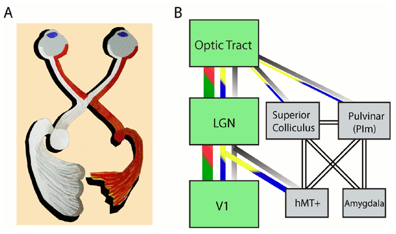
Figure 1.
(A) The major visual pathway from the eyes to the visual cortex and the reconfiguration at the optic chiasm. The right geniculostriate projection (red) is damaged and hence the lateral geniculate nucleus (LGN) is reduced in size relative to the left intact side (white). (B) Several visual pathways from the optic tract. The major pathway via the LGN to primary visual cortex (V1) is shown in green. The three main classes of retinal ganglion cell are indicated by the red-green (P-cells), gray (M-cells), and blue-yellow (non-M–non-P cells) lines. No assumptions are made about the origins of the connections indicated with the unfilled lines. Illustration in A courtesy of Betina Ip.

