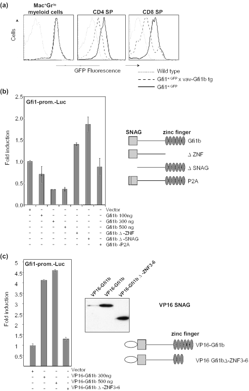
Figure 7.
Gfi1b/Gfi1 cross-regulation in bone marrow and thymocytes. (a) Bone marrow cells were stained with α-Mac1-PercpCy5.5/α-Gr1-PE. Thymocytes of Gfi1:GFP knock-in mice (Gfi1+/GFP) were stained with α-CD4-PE/α-CD8-PercpCy5.5. GFP fluorescence of gated cells was analyzed by flow cytometry as described previously (8). Histograms show the GFP fluorescence intensity of Mac1+/Gr1lo bone marrow cells, or CD4 single positive or CD8 single positiveT-lymphocytes from wild type, Gfi1+/GFP and Gfi1+/GFP/vav-Gfi1b transgenic mice as indicated. (b) A Gfi1-promoter driven luciferase construct that was described previously (8) was cotransfected into NIH3T3 cells with pCDNA3 as control and pCDNA3-expression plasmids for full-length or mutated Gfi1b as indicated. Transfection efficiency was controlled by cotransfection of beta-galactosidase expression plasmids. Fold inductions were derived by dividing the normalized luciferase activity of each value by the average of the vector control. (c) A Gfi1-promoter driven luciferase construct was cotransfected into NIH3T3 cells with pCDNA3 as control and pCDNA3-expression plasmids encoding full-length or mutated Gfi1b fused in-frame to an N-terminal VP16 AD. Fold inductions were derived as described in (b). Right panel: the expression of the VP16-Gfi1b proteins was detected by western blot using an α-VP16 antibody. Also given is a schematic representation of the VP16 fusion constructs used.

