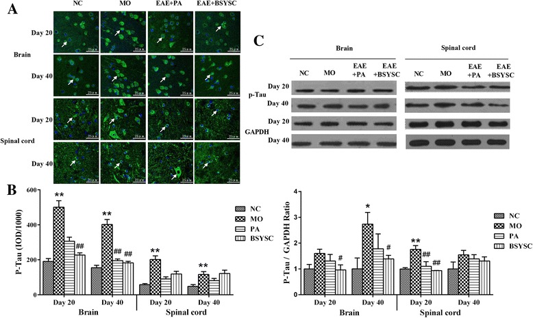
Fig. 2.

P-Tau protein expression in the brain and spinal cord of mice treated with or without BSYSC 20 and 40 dpi. The protein levels of p-Tau were reduced to different degrees in BSYSC group. a The representative immunofluorescent staining of p-Tau was shown in each group, the white arrows indicate the positive regions, magnification (×40). b Semi-quantification of integrated optical density (IOD) on p-Tau expression was exhibited. c The protein level of P-Tau was measured by Western blot and quantified using GAPDH as the loading controls. Data are expressed as means ± SE, n = 5 for each group. * p < 0.05, ** p < 0.01 vs. NC; # p < 0.05, ## p < 0.01 vs. MO
