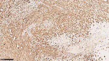
Fig. 3.

CSPG4 expression in canine osteosarcoma. Tissue samples from 29 canine osteosarcomas collected at the Diagnostic Laboratory of the Department of Animal Pathology of the University of Turin were examined. Data regarding breed, sex, age, tumor localization and clinical TNM staging were available for all dogs. The sample was fixed in 4% neutral buffered formalin, embedded in paraffin, and sectioned at 4 µm. Immunohistochemical analysis for CSPG4 was performed as previously described [74]. Briefly, sections were exposed to high-temperature antigen unmasking by incubation at 98 °C with citric acid buffer, pH 6.0. Endogenous peroxidase activity was blocked with 3% hydrogen peroxide in methanol for 30 min at room temperature. Tissue sections were incubated for 12 h at room temperature with a polyclonal anti-CSPG4 antibody (diluted 1:40, Sigma Aldrich), then 30 min with biotinylated-secondary antibody (Vectastain Elite ABC) and revealed with the ImmPACT DAB kit for peroxidase. A total score considering the proportion of positively stained tumor cells and the average staining intensity was assigned as previously described [74]. Briefly, the score indicating the positivity of tumor cells was assigned as follow: 0 (none); 1 (<1/100 or <1%); 2 (1/100–1/10 or 1–10%); 3 (1/10–1/3 or 10–30%); 4 (1/3–2/3 or 30–70%); and 5 (>2/3 or >70%). The score representing the estimated average staining intensity of positive tumor cells encompass 0 if none, 1 weak, 2 intermediate, 3 strong. The two scores were then added to each other to obtain a final score of CSPG4 expression ranging from 2 to 8. A representative image from a canine appendicular osteosarcoma is shown. Neoplastic cells are characterized by diffuse and strong cytoplasmic and membrane immunolabeling for CSPG4 and the total expression score is 8, resulted by the sum of the percentage of positive cells (=5) and the staining intensity (=3). Magnification 20X
