Fig. 5.

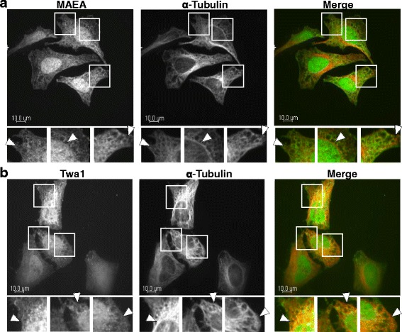
CTLH components associate with microtubules. a Hela cells were fixed and incubated with antibodies against MAEA and α-tubulin. Shown are single plane confocal images. Insets are enlarged images of the boxed regions from the above panels and arrows indicate areas of colocalization. The right panels show merged images (MAEA, green; α-tubulin, red) Scale bar: 10 μm. b Hela cells were fixed and incubated with antibodies against Twa1 and α-tubulin and analysis was performed as described above (Twa1, green; α-tubulin, red)
