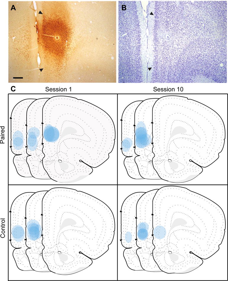
Figure 2.

Fluoro-Gold (FG) injection sites in the prelimbic cortex (PL). A photomicrograph of a representative FG injection in the PL (A) with adjacent thionin-stained section (B) used to demarcate PL borders based on a rat atlas (Swanson, 2004). Illustration of all FG injections in the PL for each training group shown on modified Swanson atlas templates (atlas Levels 6, 7 and 8; +4.2, +3.6, and +3.2mm from bregma respectively; C). Scale bar = 100 μm.
