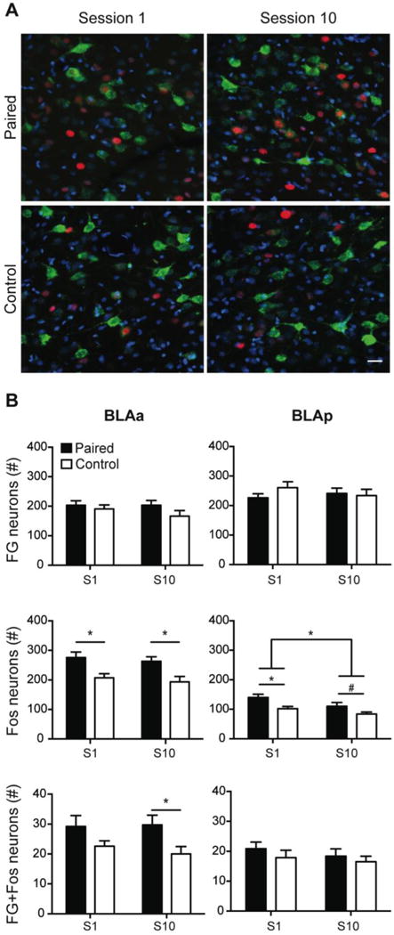
Figure 3.

Fos induction in BLA-PL projecting neurons during early and late cue-food learning. Representative images from the BLAa from each training group depicting FG-positive neurons (green), Fos-positive neurons (red), and DAPI, a nuclear counterstain (blue). Scale bar = 25μm (A). Total number of FG-positive neurons, Fos-positive neurons, and double-labeled (FG+Fos) neurons (mean ± SEM) during the first (Session 1; S1) and last (Session 10; S10) training sessions in the BLAa (left) and BLAp (right; B). *P<0.05; #P=0.087.
