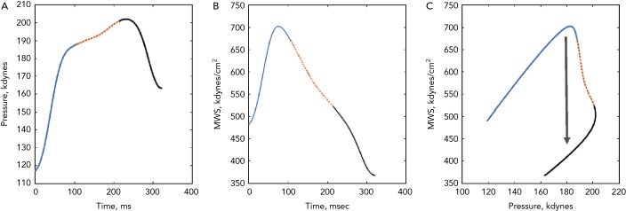
Figure 4: Time-Resolved Myocardial Wall Stress of a Normal Aged Left Ventricle.
(A) The ejection-phase aortic pressure profile. (B) The time-resolved ejection-phase myocardial wall stress (MWS). (C) The pressure–MWS relation. In all plots, the 1st, 2nd and 3rd thirds of ejection are plotted in solid blue, dotted orange, and solid black, respectively. It can be seen that MWS peaks in early systole and subsequently decreases, even in the context of increasing pressure. This is due to a mid-systolic shift in the pressure-stress relation (black arrow) that favours lower MWS for any given pressure. This shift is due to the geometric reconfiguration of the left ventricle (decreased cavity volume relative to left ventricular wall volume), and is impaired in the presence of reductions in left ventricular ejection fraction, concentric geometric remodelling and reduced early systolic ejection (reduced early-phase ejection fraction).

