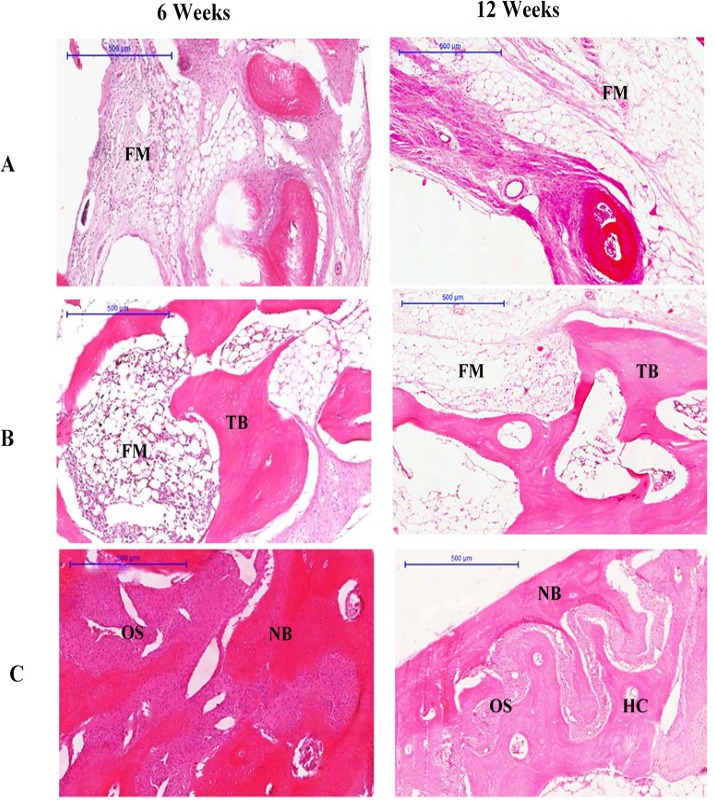
Figure 2. Photomicrographs of defect sites of (A) Group I, (B) Group II (C) Group III at 6 and 12 weeks.

Sections were stained with haematoxylin and eosin. NB, new bone; OS, osteoid; FM, fatty marrow; HC, Haversian canal; TB, trabecular bone. (Scale bar represents 500 µm.)
