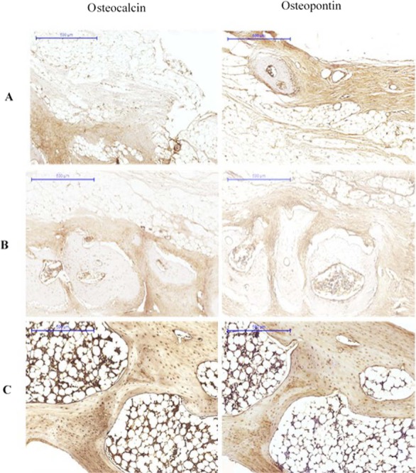
Figure 4. Immunohistological results at 12 weeks for (A) Group I, (B) Group II (C) Group III.

The pictures are arranged by staining technique (columns) and by the investigated treatment (rows). Areas that stained positive for osteocalcin and osteopontin are indicated by red arrowheads. NB, new bone; OS, osteoid; FM, fatty marrow; CB, compact bone; CAB, cancellous bone.
