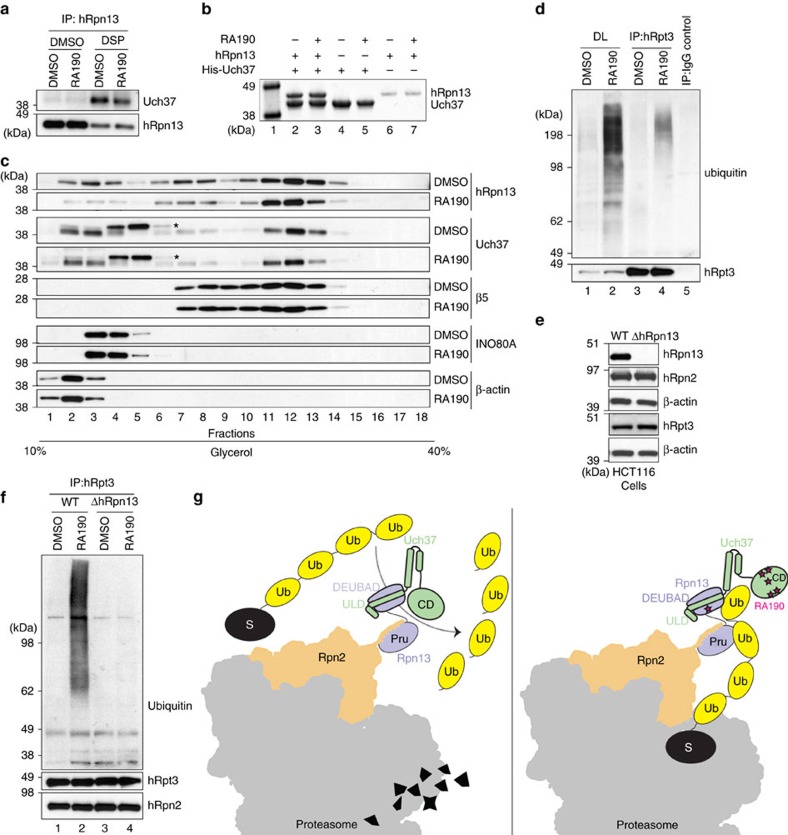
Figure 5. RA190 treatment leads to accumulation of ubiquitinated species at the proteasome.
(a) HCT116 cells were treated with 1 μM RA190 or DMSO for 24 h and then further treated with the crosslinker DSP or DMSO as a control. Cell lysates were immunoprecipitated with anti-hRpn13 antibodies and immunoprobed with anti-Uch37 antibodies. (b) His-Uch37, hRpn13 or equimolar mixture of these two proteins at 2 μM were incubated with 20-fold molar excess RA190 or DMSO and then Talon resin. Retention of hRpn13 on the Uch37-bound resin was assessed by SDS–PAGE with Coomassie staining. Protein markers are included in lane 1. (c) HCT116 cell lysates treated with 1 μM RA190 or DMSO (as a control) for 24 h were loaded onto a 10–40% linear glycerol gradient and subjected to ultracentrifugation. Gradient fractions were resolved by SDS–PAGE and immunoprobed with indicated antibodies. *Nonspecific band from the Uch37 antibody. (d) HCT116 cells were treated with 1 μM RA190 or DMSO control for 24 h and lysates immunoprecipitated with anti-hRpt3 antibodies, followed by immunoblotting as indicated. Direct loads (DLs) were included as well as an IgG control for the DMSO-treated cells. (e) Wild-type (WT) or hRpn13-deleted (ΔhRpn13) HCT116 cells were collected, lysed and immunoprobed as indicated, with β-actin as a loading control. (f) HCT116 WT and ΔhRpn13 cells were treated with 1 μM RA190 or DMSO control for 24 h and lysates immunoprecipitated with anti-hRpt3 antibodies, followed by immunoblotting as indicated. hRpn2 was used as a control to confirm immunoprecipitation of proteasome subunits. (g) Proposed mechanism of action for RA190 at the proteasome. RA190 (pink star) targets the hRpn13 DEUBAD domain without abrogating hRpn13 (periwinkle blue) interaction with ubiquitin chains (yellow) or Uch37 (green). RA190 also targets and inactivates the Uch37 catalytic domain (CD), impairing ubiquitin chain disassembly and in turn ubiquitin release at hRpn13. A portion of the 26S proteasome is represented in grey with Rpn2 in orange and substrate in black.

