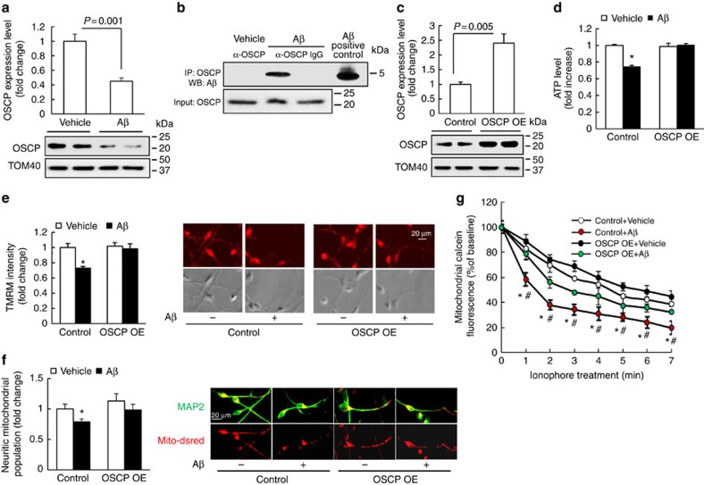
Figure 8. OSCP overexpression ameliorates Aβ-induced mitochondrial dysfunction in human neurons.
(a) Human neurons were exposed to 500 nM oligomeric Aβ1–42 for 4 days, then subjected to immunoblotting detection of OSCP levels. Aβ induced a significant reduction in OSCP expression level. The lower panel shows representative immunoblotting images. Tom40 was used as the loading control. n=4–5 samples of each group. (b) Co-immunoprecipitation of OSCP and Aβ in Aβ-treated human neurons. (c) Significantly increased OSCP expression in the OSCP overexpressing neurons. The lower panel shows representative immunoblotting images. Tom40 was used as the loading control. n=4 samples of each group. (d) Preserved ATP production by OSCP overexpression. *P<0.01 versus other groups. n=6–10 samples of each group. (e) Preserved mitochondrial membrane potential by OSCP overexpression. *P<0.01 versus other groups. n=17–23 neurons from at least 3 time independent experiments. Right panels are representative images of TMRM staining (upper row) and phase contrast (lower row). Scale bar, 20 μm. (f) Preserved neuritic mitochondrial population by OSCP OE. *P<0.05 versus other groups. Right panels are representative images. Dendrites were determined by the staining of MAP2 (green); and mitochondria were identified by mito-Dsred (red). Scale bar, 20 μm. (g) Attenuated Aβ-sensitized mPTP formation by OSCP overexpression. Ionophore (2 μM) was used to trigger mPTP formation. *P<0.05 versus vehicle-treated control and OSCP OE neurons. #P<0.05 versus Aβ-treated OSCP OE neurons. n=4–7 independent experiments. Error bars represent s.e.m.

