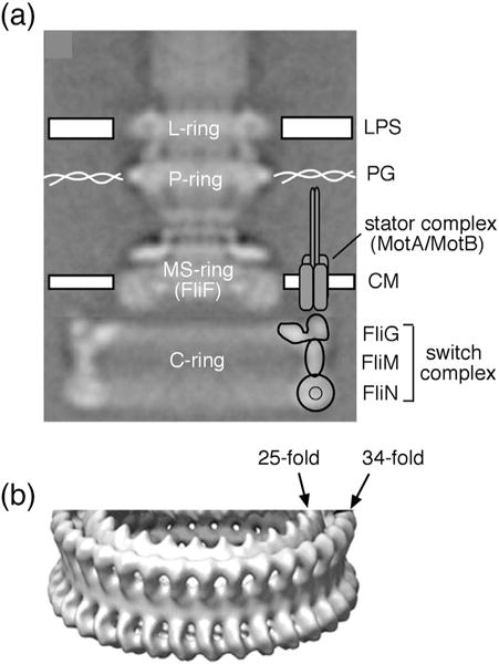
Fig. 1.

Morphology of the flagellar basal body of Salmonella. (a) EM reconstruction of the flagellar basal body from Thomas et al. [28]. The structure consists of a central rod and a set of rings. The membrane/supra-membrane (MS) ring is in and just above the cytoplasmic membrane (CM). The outer membrane (LPS) and peptidoglycan (PG) are also indicated. One stator complex is shown; motors can contain up to about a dozen independently functioning stator complexes, each attached to peptidoglycan. Approximate positions of the three switch complex proteins are indicated. (b) A tilted view of the switch complex from a higher-resolution EM reconstruction [6]. Density has been rotationally averaged to improve signal to noise. Most of the switch complex has ~34-fold symmetry, but the inner ring in the upper (membrane-proximal) part displays ~25-fold symmetry.
